It’s super effective and easy to do…
And you can do it from the comfort of your own home.

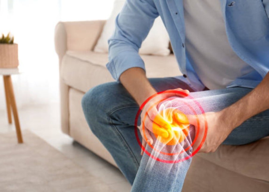
Osteoporosis is a bone disease that causes more than 8.9 million broken bones worldwide, across men and women, every year, resulting in an osteoporotic bone break every 3 seconds. Osteoporosis is a medical condition that weakens bones and makes them more likely to break. They can become so fragile that in serious cases, a strong sneeze or minor bumps can cause them to break.
Wondering why you haven’t heard more about osteoporosis? Well, one reason could be that it is known as a silent disease because you can’t see or feel bone loss, so it goes unnoticed until the first bone break occurs. Both men and women are at risk of osteoporosis. Postmenopausal women are at a higher risk after an initial fracture. These women are five times more likely to suffer another bone break within a year.
The World Health Organization, who has considered osteoporosis to be of increasing importance, many women remain untreated within six months after an osteoporosis related fracture. It’s important to be aware of the risk factors associated with osteoporosis in order to understand if you could be affected. Here are a few to consider. Previous bone break as an adult advanced age low bone mineral density parental history of hip fracture low body weight smoking.
You’ve likely heard that vitamin D, calcium, and weight bearing exercise can help keep bones healthy. It’s true. But for people with osteoporosis, it may not be enough to protect against a fracture. Osteoporosis and broken bones are not a normal part of aging. If you are a postmenopausal woman, you may be at risk for osteoporosis. It’s important to talk to your doctor about a bone health plan.
What are the common risk factors and pathophysiology associated with osteoporosis? What is the common complications associated with osteoporosis? And then finally, very briefly, we’ll look at how is it diagnosed? How is it treated? So osteoporosis is a term, it’s a compound word where osteo refers to bone porosis, pores. Simply, it just means our bones become weakened.
Let’s have a quick look at this particular diagram. It’s a typical bone, and with all bones, they have an outer covering. This outer covering is known as compact bone. This compact bone is very dense bone tissue, which means it’s strong, provides structural integrity, but most importantly, it stops compressive forces. Now, deeper to compact bone, which you can see in red, is spongy bone.
This is less dense. It has almost these little holes all the way through it, which makes it look like a sponge. And this is why it’s called spongy bone. But this tissue also allows for flexibility and strength, but without the weight associated, or as we found with a compact bone. Now, when we look at bone, I kind of think of it like a brick house. We kind of think it’s made, it’s there, it’s providing the support.
It doesn’t do much else, but in fact, it’s a very dynamic tissue, which means it’s constantly being turned over. This term is known as remodeling. And actually, this bone would probably remodel between four to ten years. So the whole thing is reabsorbed and added back between four to ten years. This is known as remodeling, which is a balancing act between taking bone away, which we call bone resorption, and adding new bone.
Now, the cells that take bone away is they’re known as osteoclasts. Now, these are kind of like macrophages that take, that eat away and take bone or reabsorb bone, whereas bone cells that add new bone are known as osteoblasts. So it’s a balancing act between these two to keep this bone remodeling process going. Now, in our early ages of life, so child adolescence, the actual osteoblasts win.
So we see more bone being formed, which actually leads to the bone mass or our bone mass being peaked. So we call this peak bone mass at the ages of about 20 to 29 years. So that means we have the strongest bones in this age. So after this age, peak bone max starts to drop off, and we actually see approximately zero, 7% drop in away each year. That means the osteoclasts become more active after the 29 years in comparison to the osteoblasts.
So what kind of things in the early phases allow the osteoblast to be more active? Well, probably the most important one are hormones. So the hormones that we see that have the greatest effect on osteoblast activity would be oestrogens, more dominant in females, testosterone more so in males, and growth hormone for both. So these hormones, which are higher in these periods, would allow this cell to be more active in that phase.
Other things is diet, specifically calcium. So calcium, high amounts of calcium will activate greater activity for the osteoblasts. And remember, vitamin D increases calcium. So those two kind of go hand in hand. So diet is also important for that peak bone mass. Other things that are important is physical activity, specifically loading physical activities, like putting your bones under stress, like resistance training.
So this is an important physical activity to generate the osteoblast activity. Now, these things feed into the peak bone mass. So if we don’t have these things working or it’s slightly reduced, we actually see we have a lower peak bone mass, which then would predispose someone to osteoporosis. Now, with the peak bone mass, because of the hormonal difference, males will have slightly higher peak bone mass than females.
So that’s important to know why females will be at higher risk of osteoporosis. It’s actually at five to one. So five females to one male. And that’s probably due largely because of the hormone. Now, other risk factors that come into leading to osteoporosis, I’m going to discuss now. But before I do that, it’s just important to note there are two types of osteoporosis.
We have primary osteoporosis and secondary osteoporosis. Primary osteoporosis is broken into further two parts. We have age related or senile osteoporosis and sex related or postmenopausal osteoporosis. And we’ll talk about that when we talk about the hormones in a second. The other type of osteoporosis is secondary. That means you have a disease, and osteoporosis is secondary to this disease. Now, some of the causes of this would be drugs.
So the most common drugs that can lead to osteoporosis would be corticosteroids, proton pump inhibitors, some antiseizure medications and heparin, they’re the most common. Smoking and alcohol also can lead to that as well. Other causes of secondary osteoporosis would be endocrinal disorders, like too much cortisol, too much thyroid hormone, too much parathyroid hormone, or a drop in some of the gonadal hormones, like oestrogen, testosterone.
Some cancers can also lead to osteoporosis, like multiple myelomas. But we’re going to focus on for the rest of this lecture on specifically the age related or senile and postmenopausal, which are the primary causes. So now let’s look at how these risk factors feed into the pathophysiology of osteoporosis, starting with age, as we said, after the age of 29 years, the osteoblast activity starts to drop in relative to the osteoclass.
So this becomes more dominant. So age, what will happen is age effects is the osteoblast number and activity will start to drop away. That means bone formation will start to decrease. Another big factor which is important is oestrogen levels, and this is for the primary osteoporosis being postmenopausal osteoporosis. So oestrogen actually has a trophic effect to osteoplasts, but also increase in or hold in number.
So as oestrogen starts to drop away after menopause, the actual effect of this will start to diminish to a point where we lose 2% of compact bone per year and 9% of spongy bone per year. And this is why up to 40% of all females will have some degree of osteoporosis. So oestrogen having a huge effect for osteoblasts, but also as the oestrogen effect starts to drop away, low oestrogen will actually cause the osteoclass activity to start increasing.
So that would be age and the sex effect. Physical activity will also see a drop in physical activity as we age. So a drop in physical activity will, will actually make the osteoblast activity less. Therefore, we have a decrease in bone formation. So that’s the physical activity. Diet or diet, this would be calcium and vitamin D. Also. These things start to diminish in our diets as we age. So that means bone formation also decrease. Drugs we spoke about.
But I’ll just add one important one. Corticosteroids, which we see used for decreasing the immune response in some autoimmune conditions or asthma, et cetera. So corticosteroids would actually increase the activity of osteoclast. That means we have an increase in bone reabsorption and then finally, genetics. Well, this goes back to the peak bone mass in our earlier phases of life.
So it seems that Caucasians and Asians have a lower peak bone mass compared to, say, Africans. So that’s important for predisposing one to osteoporosis. So from a decrease in bone formation and an increase reabsorption as we get older, what that will do is decrease bone mass. And by doing that, we have a decrease in bone strength.
Now, what happens is we start to see greater holes or spaces, particularly in the medullary bone or the spongy bone, which is why it’s called parosis. So this, with a thinning of the compact bone, is the most common changes that we see histologically in bones with osteoporosis.
So this is then going to predispose us to fractures. The most common fracture location is the vertebra. Second to that is the neck of femur. Then we see wrist and ribs thereafter. Now, it’s important to note that the diagnosis for osteoporosis is where we have a reduction in peak bone mass by at least 2.5 standard
One in three women and one in five men aged 50 or over suffer from osteoporosis, which can lead to bone fractures. The disease makes bones weak and brittle. And the International Osteoporosis foundation says that 9 million fractures occur annually, putting a heavy burden not only on patients, but also their families. Japanese researchers are trying to determine the mechanism behind osteoporosis at a cellular level to find a new treatment.
This image shows a fracture in the spine of an osteoporosis patient. One of the bones was crushed under the patient’s weight. Osteoporosis is often called a silent disease because it progresses without symptoms until one day a sneeze or cough can result in a bone fracture. Erika set out to learn how doctors diagnose this symptomless condition.
Takayuki hosoi has spent many years treating osteoporosis. So, Dr. So how do you actually diagnose osteoporosis? Great. I’m looking forward to trying it. OSOi measures bone density with a special device using x rays. Erica is in her fifty s. This is said to be the age at which bone density starts to decrease rapidly in women. The test takes about minutes.
Kariga kyono kede hi. Masna hidariga. Yoti migiga hidari. No. Cocaine also compares bone density in her lower spine and upper thigh bone with the averages of those areas for younger people. So it’s quite good. Very good. Very good. Oh, good. The benchmark values for bone density are the averages for 20 to 44 year olds in the lower spine and for 20 to 29 year olds in the upper thigh. Bone readings that are 70% of these benchmarks or less are diagnosed as osteoporosis. The density of a bone depends on its internal structure.
There are countless pillars of calcium inside a bone. Their compactness is what is referred to as bone density. Cells called osteoclasts are located on the surface of these pillars. They release acid and enzymes to melt the pillars. You other cells, called osteoblasts, deposit calcium and protein into the cavities created by the osteoclasts. In this way, the bones constantly undergo a metabolic process.
Dr. Ho so the bone is constantly turning over. What’s the purpose of this? Kara honey. No. Kyodo Tamas Tabiniwa, kono tanoba, James Marimas. Amazing. When you think about it, cells called osteocytes, located inside the pillars of calcium, play a key role in bone metabolism. The osteocytes are affected by the female hormone estrogen estrogen controls the osteocytes and gets them to suppress the excessive destruction of pillars by osteoclasts and to promote bone formation by osteoblasts.
However, estrogen levels in women begin to fall a few years before menopause. When this happens, the osteoblasts can no longer keep up with the osteoclasts resulting in a gradual decrease in bone density in men. Male hormones which are involved in the maintenance and building of muscle are converted to estrogen. Dancing about you are kiruge hormone kankyo karakoto ninejokoto ari mascadamo or konan kino yoni stone sagar koto and I kuriga dana Okinachi guide Jose and Dr. Hussein at the moment, what sort of treatments are available for osteoporosis?
Mauiku Akimaski or kyodokuni osiri say oh hagimask city succinct animals the Konakada chiva many so Dr. Hossa what are some of the side effects of these drug therapies? Hakosaibo do you know? Motomoto honeymoon toky dustini hostage so marindasara decimal take a but decimal the yona kotoga animals at Miyaku Nada senior kidney kuno our bones are maintained by the coordinated efforts of the cells inside them.
Scientists had known this but had been unable to observe these cells at workload. When bones are taken out of the body, the cells inside them die. It was considered impossible to see the cells while they were living. However, one researcher successfully captured images showing the activities of living bone cells. He even developed a special microscope.
Masaru Ishi is an immunologist at Osaka University Medical School in western Japan. Kononakani modo soyano matoka beto he sokoni matato maneu mitoka atatakai ishi anesthesizes a mouse and puts it on a warm plate.
The device maintains a temperature that allows the animal’s cells to function as usual. The system also monitors the mouse’s pulse to ensure it’s not overly anesthesized when observing its skull, he removes a tiny section of skin on its head. Otherwise, the mouse is kept in as normal a state as possible. Possible bone is made from hard tissue, which is opaque.
The cells inside cannot be seen with a conventional microscope. Ishi attempted to view them using a near infrared laser, for which absorption and reflection differ depending on the material. He made repeated subtle adjustments to the laser’s wavelength and intensity. Photo Science show them so the music stands man Ishi finally succeeded in capturing a view of live osteoclasts for the first time in the world.
Genkato toma and or taskai so it was in 2016 that Ishi managed to capture the world’s first image of osteoclasts in action. The blue area is bone. The green parts are the osteoclasts. The osteoclasts are releasing a red substance. It’s the acid that melts calcium. The image of live osteoclasts melting bone tissue appeared in an international science journal, surprising the world.
You umasan just the Koniko Sango dashiru so ketteri shinkan dot to go the Doklas katigoto mita in a kataras no. There bakana is. Two years later, in 2018, Ishi also succeeded in capturing osteoclasts and osteoblasts coming into direct contact with each other. The red objects are osteoclasts. The light blue ones are osteoblasts. This revealed that the osteoclasts do not melt bone while in contact with osteoblasts.
The two groups of cells are thought to communicate with each other to maintain bone homeostasis. Ishi became fascinated with intravital imaging while doing research in the US at the National Institute of Allergy and Infectious Diseases. As a clinical doctor, he’d been treating patients suffering from bone diseases. He aimed to learn the true cause of their conditions and apply his findings to treatment. In 2019, he finally discovered the culprit.
It turned out to be a type of cell called a malignant osteoclast, which destroys bone tissue excessively. Hako saibo isuri there sujo noi hakosai boga mahatara kisuite these images show the activities of normal and malignant osteoclasts. The red objects are the osteoclasts. The acid they release is shown in yellow, normal osteoclasts release only small amounts of acid.
Malignant osteoclasts secrete far more. Until Ishi’s discovery, all osteoclasts were believed to originate from cells called osteoclast precursors. Ishi found that malignant osteoclasts come from completely different and unusual precursor cells. In other words, the two types of osteoclasts originate from different kinds of cells.
Shikamo the snehonyo, kwasuto kono bioti boa kasushi zuto kwasiru you ishi suspected that if he could prevent the cell differentiation that resulted in malignant osteoclasts, bone diseases could be cured. Detailed analyses revealed that the cells that turn into malignant osteoclasts are affected by a substance called fox M one. Ishi conducted an experiment in which he injected a fox m one inhibitor into mice.
Bone destruction progressed in the mice that weren’t given the injection. However, it was suppressed in those that were given the inhibitor. Ishi believes that the discovery could be used to prevent osteoporosis. Jikanimotima so, guys, in Japan, many researchers are looking at lifestyle improvements as a way to prevent osteoporosis. Yoshiko Ishimi is a pharmacist specializing in nutritional science.
She has been studying food components called isoflavones contained in soybeans and their effects on osteoporosis risk reduction. And how actually are the soy isoflavones? How do they prevent osteoporosis? Ano dijon haitirua jose hodmoni, nitarun desuna the design of the honeno or kenko iji, sternokangai maso when soy isoflavones reach the intestines, gut bacteria may transform them into a substance called equal.
Equal is very similar in molecular structure to estrogen, the female hormone. It is considered highly effective in preventing osteoporosis. Does everyone have this gut bacteria to convert to transform these soy isoflavoins to equal.
Sono sayo no kata obey no katawa sanju passenger hoga daiso yori oku manichi mason kangari maso so what happens if you cannot make a call? To see how soy isoflavones affect bone density in people who cannot produce equal, ishimi conducted an experiment on japanese women within five years after reaching menopause.
When those who can’t generate equal consumed soy isoflavones contained in 200 grams of tofu daily, their bone density fell 2% in one year. In people who didn’t consume soy isoflavones, bone density dropped about 5%. And professor, what sort of foods contain soil isoflavoins? Itchiba kinakodesna daiso sonomamatsobustamono descara kinakodeska tonyudestoka natomos higashi no ho nostota nishino higashi noho nosto tatino shima chigaga nato.
There. Nato kenkoni kangai maso. So obviously Nato is something that’s never going to become popular overseas. Don’t worry, erica. There’s a fruit that boosts the effect of soy isoflavones when eaten together. Ishimi conducted a joint study with a university in New Zealand. They looked at how consuming soy isoflavones and kiwi together affected the bones of women in the country who were within five years after reaching menopause.
The results showed the marker related to the risk of bone fractures was much lower in people who ate both than in those who consumed soy isoflavums only. Jisani tobutoji kendeva gurin kiwito go to the kiwi toyoho shira vitamin k no gayuri kiwi no naka no vitamin kono tai kanki stemas no that surrega sayo stem no kanato. That’s interesting. So, kiwi. I think there’ll be a boom with kiwi.
So, professor, can diet alone prevent osteoporosis? So I can’t just have my morning smoothie with soy milk and then kiwi fruit and then expect strong bones if I’m sitting in front of my computer every day. Thank you so much. Now, Hosoi, the osteoporosis expert will explain what kinds of exercise can make the bones stronger. Mitsno pointo garimas.
Applying impact and weight to the bones and exercising the muscles near them stimulates osteocytes, the cells that play a key role in bone metabolism. The bones become stronger as a result. The first exercise, called the heel drop will jolt your bones. It is strictly preventive as it can cause fractures in people who already have osteoporosis.
You umasaki tasio niste agate don’t touch kara homony as no honeymo sorry masnode kahaniki sri kangai mas you you do three sets of ten repetitions a day. This is a really easy exercise to do. Next is a weight bearing exercise. Place your hand on the back of a chair, lift your knee and stand still for 1 minute. If you prefer, you can use both hands for better support.
Katashu agamas Taksangiri Masa Society can anyone do this exercise? Tomato nikai saga katari sarah kosa so sagatamani also, a minute’s a long time. I know when you start off in the exit it’s quite a long time. It’s a powerful exercise. Next is squats to strengthen the lower body muscles and apply weight to the bones. When you bend your legs, make sure your knees don’t stick out beyond your toes.
If your knees hurt or if you have difficulty maintaining balance, you will still benefit from repeatedly sitting and standing from a chair. Isn’t it koskaki to your knee. Stay Osiri auto stick Oscatawa Naito Adamasan Kiniko Scalkoto attend to yobo Nagarukoto Kinika shirukotoka konani tamiyo motoras so why then is it important first not to put the knees too far forward.
Kizagama toe scalkinikuga kunakunagima mandusi Maskono Kinik Mask Sunday mata it sunbirita hoga coca garima aim to do three sets of ten repetitions a day. Because I think when you’re doing it slowly, you can really feel it more. I think so many people do squats so quickly. This is because you’re really focused on these muscles. It’s finally we’ll show you the correct form for walking, which applies both impact and load to the bones.
Tarasku arakoto kenkonimoditisanaikoto if you walk with your gaze lowered and your back hunched, your feet won’t touch the ground heels first. This will not distribute the weight on your bones properly. Say konarimas das karito katrio kakir koto they always kari you see running through kotokotonio tencomas kencot johans you know.
Karamega mario kinikini okina ako State Thai sama ted Thai kang mo kitai nag para because doctor, some of these exercises look deceptively easy, but when you actually do them they’re quite powerful, and I could really feel it. I think it’s great for balance as well, but I think everyone watching is going to be so inspired to do them to help keep our bones nice and healthy and strong. So, doc, thank you so much for your time today.
About the osteoporosis treatment guidelines issued by the ACP, the American College of Physicians. The focus of the guidelines is on treatment, but before I begin, I do want to discuss screening. Last weighed in on by the USPSTF, the United States Preventive Service Task Force, in 2018.
Note those guidelines are being updated. What they say is that osteoporosis should be screened for in women older than 65 years of age and in those who are younger who are at increased risk. Based on a risk assessment tool, usually the FRACS tool, they state that there’s not enough evidence to weigh in for or against screening men.
The other large organization that weighs in on screening, the bone Health and Osteoporosis foundation, agrees with the USPSTF, but in addition says that we should be screening men over 70 and men who are younger, 50 to 69, who have risk factors. In addition, consider screening or you should screen anyone who has a fracture after low impact or no trauma.
Let’s now go on to the ACP guidelines. Osteoporosis is defined as bone mineral density at the femoral neck or the lumber spine, or both, with a t score less than negative two and a half. Recommendations are as follows. For postmenopausal women with osteoporosis, you should use a bisphosphonate as first line treatment in order to reduce the risk of future fractures.
This is given a strong recommendation based on a high certainty of evidence. This phosphonates versus placebo over three years leads to one fewer hip fracture per 150 patients treated and one fewer vertebral fracture per 50 people treated. All the other recommendations in the guidelines are considered quote conditional, unquote. Conditional recommendation is one that most people would do, but there are individuals who might not do that based on level of risk and personal preference.
For men, for instance, treatment of osteoporosis is given a conditional recommendation not because the evidence suggests that it’s not as good, but just because there’s not as much evidence there. If you are to treat a male with an osteoporosis with osteoporosis, it should be initial treatment using abyss phosphonase.
Again, remember, men do get osteoporosis and account for about 30% of hip fractures. This is not a surprise to anyone who takes care of older adults. For postmenopausal women or men who you would want to treat but who can’t tolerate a bisphosphonate, then the recommendations are to use denasumab and that can be used as second line treatment to reduce the risk of fractures. Remember, bisphosphonates and denasumab are antiresorptive drugs, meaning they slow the progression of osteoporosis.
The anabolic drugs, on the other hand, ramasumab and teraparitide increase bone density. The anabolic agents should only be used in women with primary osteoporosis who are at very high risk of fractures. And use of these agents always needs to be followed by then using an antiresorptive agent because otherwise there’s a risk of rebound osteoporosis and an increased risk of vertebral fractures. Now how about osteopenia, which a lot of people have?
The guidelines recommend that for women over 65 with osteopenia we ought to use an individualized approach influenced by the level of risk as we sort out their other risk factors, including things like increased age, female sex, low body rate, current smoking, hip fracture in a parent, fall risk and a personal history of fracture.
The guidelines note that increasing the duration of bisphosphonate therapy longer than three to five years does reduce the risk of additional vertebral fractures, but it doesn’t affect the risk of other fractures and it increases the risk of osteo necrosis of the jaw and atypical hip fractures. Therefore, most of the time we ought to be using bisphosphonates only for three to five years, unless someone is at extremely high risk.
It’s also important to note that there’s a five fold higher risk for atypical femoral fractures among asian women. Of course, don’t forget about vitamin D, don’t forget about calcium, and most importantly, don’t forget about exercise, particularly exercise aimed at improving balance and quadricep strength, which helps prevent falls. I’m interested in your thoughts on this. Go to Home