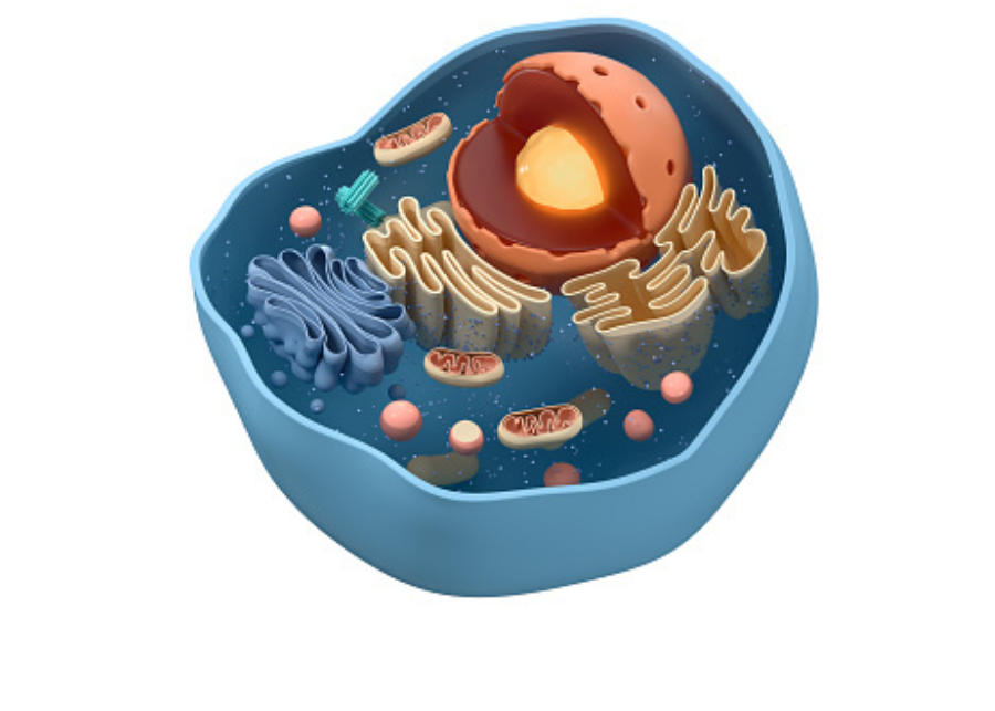
All living organisms are composed of cells. Cells are responsible for all anatomical and physiological features of all body systems. Different cell types can vary greatly in shape and size, but they all have a common structure and similar components. A typical cell cell is enclosed in a plasma membrane and contains a nucleus and a cytoplasm. The plasma membrane serves as the cell’s boundary, controlling the traffic of substances in and out of the cell.
It is also the site of communication between the cell and its environment. The membrane consists mainly of two layers of phospholipids with their hydrophilic heads, the phosphate groups facing the aqueous environments inside and outside the cell, and their hydrophobic fatty acid tails facing in together. Other membrane lipids include cholesterol, which is essential to membrane structure and fluidity, and glycolipids, which maintain membrane stability and facilitate cell to cell interactions.
The lipid membrane is dotted with membrane proteins, of which there are two types, integral or transmembrane proteins, which span across the membrane, some passing through multiple times. Some transmembrane proteins have a small carbohydrate chain on the outside of the cell and peripheral proteins, which attach to the membrane on the inside. A peripheral protein typically functions together with an integral protein.
Membrane proteins fulfill a variety of functions. As receptors or receptorassociated proteins. They receive messages from outside the cell. For example, a nonsteroid hormone must bind to a membrane receptor and act via several other membrane proteins to activate a cellular response. Each receptor is specific to a certain molecule. As ion channels or transport proteins, they help move charged particles and large, uncharged polar molecules across the cell membrane.
As adhesion molecules, they help cells adhere to each other and to the extracellular matrix. As enzymes, they catalyze reactions that are required outside the cell but in the vicinity of the cell membrane. Transmembrane glycoproteins also serve as surface antigens, determining the cell’s identity on top of the cell membrane. Some cells have surface extensions that carry out specialized functions.
Examples include microvilli that increase the surface area in the small intestine cilia that move mucus in the respiratory tract, and flagella that are responsible for the movements of sperm cells. The nucleus contains genetic material, the DNA, and is where DNA replication and transcription, the major step of gene expression, take place. Most cells have one nucleus, with the exception of red blood cells, which have none, and some other cells that have multiple nuclei.
The nuclear envelope surrounding the nucleus consists of two membranes, inner and outer, each of which is a phospholipid bilayer. The envelope is dotted with nuclear pores protein complexes that provide controlled passage between the nucleus and cytoplasm chromosomes are strands of DNA wrapped around proteins. Under a light microscope. Chromosomes are only visible during cell division when they are highly condensed.
Instead, the most prominent feature of the nucleus is the nucleolus, the area around the clusters of ribosomal rna genes. This is where ribosomal rnas are made and where ribosomes are assembled. Ribosome then move to the cytoplasm to fulfill their function in protein synthesis. The cytoplasm includes a gel like liquid called cytosol, various organelles and cytoskeleton, the endoplasmic reticulum, ER, Golgi apparatus, and vesicles constitute the intracellular membrane system.
The ER is a network of connected flattened sacs called cisterni. Its membrane is continuous with the outer nuclear membrane. Part of the ER appears rough, as it is covered with ribosomes. This is where the synthesis of secretary and transmembrane proteins take place. These proteins have a signal sequence within their amino terminus, which, as soon as it emerges from the ribosome, targets the rna ribosome complex to the ER membrane, where translation continues.
The emergent polypeptide enters the ER membrane as it is being translated. Transmembrane proteins, identified by the presence of a hydrophobic stretch, stay in ER membranes while secretary proteins are released into the ER. Lumen, the smooth part of the ER, synthesizes lipids and lipid components of cell membranes. As lipids are produced, they are inserted into the ER membrane. Membrane proteins, lipids and secretary proteins are then packaged into vesicles to be transported to the Golgi, where proteins undergo post translational modifications.
Vesicles pinch off from ER membranes, travel to golgi apparatus, fuse with golgi membranes, and release their content. The golgi is a stack of separated cisterni. Each contains a set of enzymes responsible for a certain step in protein maturation. Similar vesicles transport lipids and proteins from one cisterna to another and ultimately to their destinations.
The plasma membrane lysosomes or storage vesicles the destination of a protein is typically determined by a signal sequence acting as an address tag within the protein. The ER is also a major site for metabolism and storage of calcium, whose release is a trigger for many cellular processes. Lysosomes are vesicles containing hydrolases that break down macromolecules into their building units, which can then be recycled.
The enzymes are activated by the acidic environment within lysosomes. In white blood cells, lysosomes digest phagocytized bacteria and play a role in immune response. Mitochondria are best known as the cell’s powerhouses. This is where energy is extracted from food compounds and stored in energy rich molecules. A mitochondrion has two membranes.
The inner membrane has multiple folds called christi. Two of the three main steps of cellular respiration occur in the mitochondria, citric acid cycle in the matrix and oxidative phosphorylation on the christi. Cytoskeleton is a network of protein filaments that fulfill a variety of functions. There are three types of filaments, microfilaments, intermediate filaments and microtubules.
Microfilaments are made of the protein actin. They enable muscle contraction, provide support for microvilli, produce cell movements and play a role in cell division. Intermediate filaments are made of different proteins in different cells. Their roles are mostly supportive. Microtubules are large tubes of 13 protofilaments. Each is a long chain of tubulent dimers.
A centriole is a short cylinder of nine triplets of microtubules. A cell typically has two centrioles lying perpendicular to each other forming a structure called centrosome. Centrosome serves as a microtubule organizing center from which microtubules grow out into the cytoplasm. In nondividing cells, microtubule networks hold organelles in place during cell division. They form the mitotic spindle that guides chromosome movements. Microtubules are also responsible for the movements of cilia and flagella.

These adults tried a simple 10-second “NASA stretch” first thing in the morning – and their crippling back pain vanished instantly.
Cells are the smallest living units of an organism. All cells have three things in common, no matter what type of cell they are. All cells have a cell membrane, which separates the inside of the cell from its environment. It cytoplasm, which is a jelly like fluid, and DNA, which is the cell’s genetic material. There are two broad categories of cells. The first category is eukaryotic cells.
They have organelles, which include the nucleus and other special parts. Eukaryotic cells are more advanced complex cells, such as those found in plants and animals. The second category is prokaryotic cells. They don’t have a nucleus or membrane enclosed organelles. They do have genetic material, but it’s not contained within a nucleus. Prokaryotic cells are always one cell or unicellular organisms such as bacteria.
So what are organelles? Organelle means little organ. Organelles are the specialized parts of a cell that have unique jobs to perform. Let’s start with the nucleus, the control center of the cell. The nucleus contains DNA or genetic material. DNA dictates what the cell is going to do and how it’s going to do it. Chromatin is the tangled, spread out form of DNA found inside the nuclear membrane. When a cell is ready to divide, dna condenses into structures known as chromosomes.
The nucleus also contains a nucleolus, which is a structure where ribosomes are made. After ribosomes leave the nucleus, they will have the important job of synthesizing or making proteins. Outside the nucleus, the ribosomes and the rest of the organelles float around in cytoplasm, which is the jelly like substance. Ribosomes may wander freely within the cytoplasm or attach to the endoplasmic reticulum, sometimes abbreviated as ER. There are two types of ER.
Rough ER has ribosomes attached to it, and smooth ER doesn’t have ribosomes attached to it. The endoplasmic reticulum is a membrane enclosed passageway for transporting materials, such as the proteins synthesized by ribosomes. Proteins and other materials emerge from the endoplasmic reticulum in small vesicles, where the golgi apparatus, sometimes called the golgi body, receives them. As proteins move through the Golgi body, they’re customized into forms that the cell can use.
The Golgi body does this by folding the proteins into usable shapes or adding other materials onto them, such as lipids or carbohydrates. Vacuoles are saclike structures that store different materials. Here in this plant cell, the central vacuole stores water going back to the animal cell, you will see an organelle called a lysosome. Lysosomes are the garbage collectors that take in damaged or worn out cell parts. They are filled with enzymes that break down this cellular debris.
The mitochondrion is an organelle that is the powerhouse for both animal and plant cells. During a process called cellular respiration, the mitochondria make atp molecules that provide the energy for all of the cell’s activities. Cells that need more energy have more mitochondria. Meanwhile, the cell maintains its shape through a cytoskeleton. The cytoskeleton includes the threadlike microfilaments, which are made of protein, and microtubules, which are thin, hollow tubes.
Some organisms, such as plants that are photoautotrophic, meaning they capture sunlight for energy, have cells with an organelle called a chloroplast. The chloroplast is where photosynthesis happens. It’s green because it has a green pigment called chlorophyll. Plant cells also have a cell wall outside of their cell membranes that shape, support, and protect the plant cell. Animal cells never have a cell wall. There are many other unique structures that only some cells have.
Here are just a few. In humans, for example, the respiratory tract is lined with cells that have cilia. These are microscopic, hairlike projections that can move in waves. This feature helps trap inhaled particles in the air and expels them when you cough. Another unique feature in some cells is flagella. Some bacteria have flagella. A flagellum is like a little tail that can help a cell move or propel itself. The only human cell that has a flagellum is a sperm cell.
In summary, remember, eukaryotic cells are plant and animal cells with a nucleus and membrane enclosed organelles, while prokaryotic cells are unicellular organisms. Without these things, all cells have a cell membrane, cytoplasm, and genetic material. And even though only plant cells have chloroplasts, both plant and animal cells have mitochondria.
Have you ever been sitting in class and thought to yourself, I wonder what my skin cells are doing right now at this very moment. This kind of pondering may be unique to me, maybe, but wouldn’t we at some point wonder what our cells are doing right now? Because if you remember, as part of the cell theory, you are made of cells. All of things are made of one or more cells.
Many multicellular organisms like you have cells that work together, working together as part of a body tissue, body tissues working together as part of an organ, organs working together as part of an organ system. Your cells are specialized to work in these different levels of organization. You have skin cells, stomach cells, muscle cells, just to name a few. And their functions need to be regulated. These cells actually are regulated as part of something called the cell cycle.
And that is going to relate to my question of I wonder what my cells are doing right now. Cells themselves can grow in size, but let’s put it in perspective now. A multicellular organism isn’t growing because each individual cell is getting bigger. A multicellular organism itself grows by making more cells, by the cells making more cells, by dividing. That’s cell reproduction.
One reason that you are bigger than you were when you were five, unless you are five, is because your cells have divided to make more cells. Mitosis and the cytokinesis that follows that splits a cytoplasm allows you to make new body cells. But you don’t want that cell division happening all the time. Why? It is likely that you’ve heard the term cancer before. We have had family members that have battled cancer before.
It is definitely a relevant topic for all of us. Cancer is in part due to cells that divide too frequently. The cells are not regulated. They are uncontrolled. Cancer cells can have other problems too. They might not be able to communicate with other healthy cells. They may not be able to carry out normal cell functions. They may not securely anchor themselves like other cells do, which can make them more likely to travel somewhere else.
Some cancer cells have the ability to secrete their own growth hormone. That makes blood vessels divert over to those cancer cells and supply the cancer cell with nutrients which can take nutrients away from healthy cells. Why do cancer cells become this way? Well, there is a lot of research in this area. With some cancers, there may be genetic links, making some cells more susceptible to having problems. These genetic factors might run in families.
Exposure to toxins, radiation, excessive exposure to uv light. All of these can be risk factors for some cells to become cancerous. The uncontrolled growth that cancer cells have gives rise to more cells like them, which can develop into a tumor. Some tumors stay put, but some do not. Now the good news is that scientists continue to develop better treatments, which include destroying the cancer cells with radiation or medications such as chemotherapy, which will target cells that divide frequently.
Maybe someday you will be part of helping to meet the challenge of trying to eliminate cancer, because the fact remains that these cells are not participating in the cell cycle like they should. So what is the cell cycle? The cell cycle is often represented as a pie chart. Like this, cells are either in one of two different phases, a phase called interphase, where the cells themselves are growing, replicating their dna, doing their cell functions, or they are in m phase, which includes mitosis and the actual splitting of the cytoplasm cytokinesis.
So this M phase is where cells actually divide to make more cells. But cells spend most of their time in interphase, so most of the time they’re not dividing. Now, depending on what kind of cell it might do, mitosis more or less often. For example, your hair follicle cells do mitosis frequently, which is why your hair can grow at the rate that it does. It’s also why many cancer drugs may also target hair follicle cells, because many cancer drugs go after cells that do cell division frequently.
It’s a big deal for cells to hit this mphase. If a cell has an error, a harmful mutation, for example, you do not want it to divide because then it will create another cell that has this same issue. That’s where checkpoints come in handy. Along the cell cycle, there are checkpoints to check that the cell is growing well and replicating its dna correctly and doing everything it’s supposed to do correctly before it divides. To better understand those checkpoints, let’s further divide this cell cycle pie chart. We have g one, s g two.
All three of those are part of interphase. Then we have m phase, where mitosis will happen. During g one, the cell individually itself grows. Then it replicates its dna. In s phase. You can remember that because the s is for synthesis, which means to make something and it’s making dna. Then g two, the cell grows some more in preparation for mitosis.
So let’s take a look at checkpoints. We’ve got one here. In g one, this checkpoint checks. Is the cell growing well enough? Is its dna damaged? Because if it is, you definitely don’t want it to move on to s phase, where it would replicate dna. Does the cell have the resources it needs if it were to keep moving on. This checkpoint in g two checks if the dna was replicated correctly back in s phase, is it growing well enough? Does it have the resources it needs to continue? Okay then, moving on.
This next checkpoint in mphase is my favorite checkpoint. It checks in the stage metaphase to make sure the chromosomes, which are made of dna, are lined up in the middle correctly, that they’re all attached to the spindle correctly, because if they’re not, the chromosomes will not be separated correctly. So now you may have two big questions. First, what happens if the cell doesn’t meet the requirements of the checkpoint?
And second, what is doing the regulating of this cycle anyway? To address the first question, if the reason the cell can’t go past the checkpoint is a reason that can be fixed, the cell may kind of pause here until it can fix that issue. But if it can’t be fixed, then the cell does something called apoptosis, which basically means the cell self destructs. This ensures that a cell that is damaged beyond repair will not go on to divide.
So what is doing the regulating anyway? We’ve mentioned before that proteins are a big deal. Genes in your body can code for proteins that do an assortment of functions. And there are many proteins involved with regulating the cell cycle. Some of them are positive regulators because they allow moving forward in the cycle, and some are negative regulators that might make things stop. The proteins themselves can be sensitive to cues inside and outside of the cell.
So, for example, two proteins that are involved in positive regulation are cyclin and Cdk. Cdk is specifically an enzyme protein, a fancy kind called a kinase, which is worth a google. Cdk can have different forms of cyclone protein bound to it. Different types of cyclone rise and fall throughout the cell cycle.
And the rising and falling is based on a variety of signals to determine when the cell should move on to the next cell cycle phase. Typically, each cell cycle phase, g one s g two m will tend to have a different cyclin binding with the Cdk. The rise and fall of cyclin types and the role Cdk has when it’s active is a fascinating subject to explore. Remember that vocabulary word we said, apoptosis. Proteins that are negative regulators.
For example, a protein called p 53, can be involved in initiating apoptosis. Again, we encourage you to explore beyond the video one last thing to mention. There are some cells that don’t go through the phases we mentioned because they’re actually in g zero. That’s a zero, by the way, and not a no because it was a no. Then it’d say go. And g zero is kind of the opposite of that. G zero is a resting phase. Now, cells here are still performing cell functions, but they’re not preparing to divide.
Some cells go here temporarily, maybe if there’s not enough resources around, for example. But some, like many types of neurons in your brain and spinal cord, may stay here permanently. If they stay here permanently, they’ll never get to mphase, so they will not divide. This can be one reason why a major injury to the brain or spinal cord can have challenges with healing, as many of those cells may not be able to replicate a topic that definitely continues to be researched. Well, that’s it for the Meiba sisters, and we remind you to stay curious. Go to Home